


ABDFAN Kyoto Kagaku 41900-030
Ultrasound Examination Training Model
(With Pathologies)
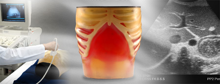
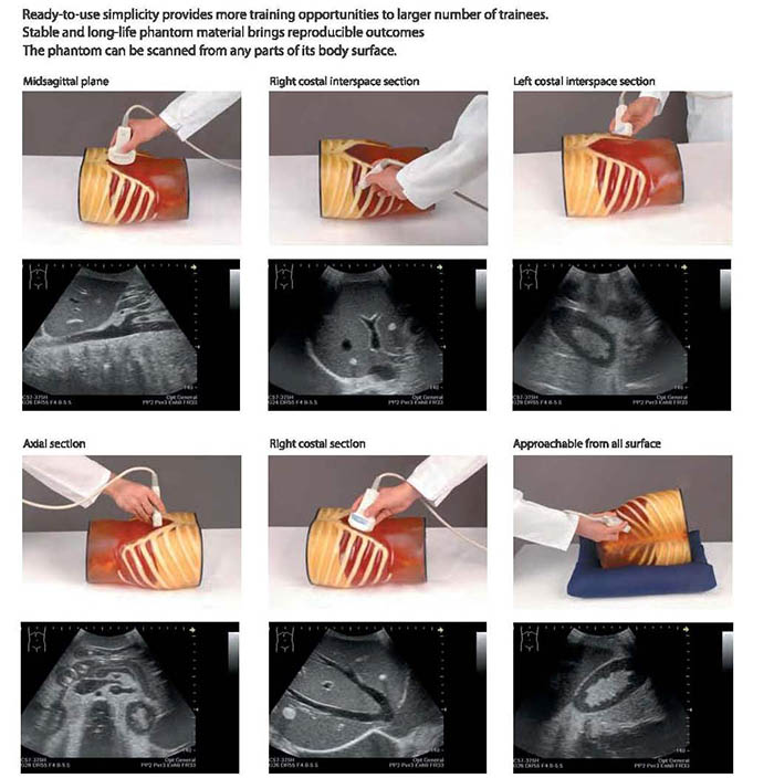
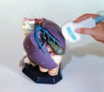
Unique high-fidelity ultrasound phantom facilitates effective training in abdominal ultrasound scanning with your own clinical devices. Simulated lesions embedded as targets provide wider educational opportunities.
- Inanimate tool for training of a novice or demonstration of technique by an expert.
- Any ultrasound device with a convex probe can be used.
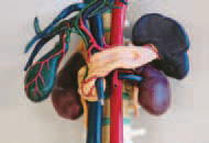
- Detailed hepatobiliary, pancreatic and other abdominal anatomy meeting requirement for excellent training.
- Eight Couinaud’s hepatic segments can be localized.
- Near real-size organs, structures and abnormal lesions.
- Durable long-life phantom materials.
Four variations of Pathologies include: Hepatic Lesions (cystic and solid), gallbladder and bile duct stones, pancreatic tumors (one invading the portal vein), splenic lesions, both kidney lesions, and left adrenal tumor.
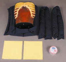
The phantom includes:
Materials: urethane elastomer |
 |
| 41900-030 |
ABDFAN Ultrasound Phantom (with pathologies) |
Includes: 1 Ultrasound phantom "ABDFAN" 2 Silicon sheets & 1 set of positioning pillows 1 Talcum powder 1 Carrying Case Size: 18 x 25 x 28 h cm, 12 kg |
| 41900-020 | ABDFAN abdominal training set |
Includes: ABDFAN set & ECHOZOU anatomical model 30 x 70 x 50 cm, 20 kg |
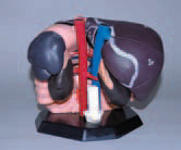
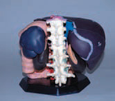
ECHOZOU Kyoto Kagaku 11224-000
Internal Organ Anatomical Model
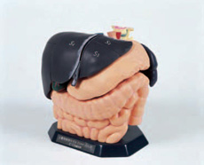
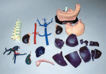
Separates into 20 parts:
- Hepatic segment S1: caudate lobe
- Hepatic segment S2: upper left lateral segment
- Hepatic segment S3: lower left lateral segment
- Hepatic segment S4: left medial segment
- Hepatic segment S5: lower right anterior segment
- Hepatic segment S6: lower right posterior segment
- Hepatic segment S7: upper right posterior segment
- Hepatic segment S8: upper right anterior segment
- Portal vein, Bile duct and Hepatic vein
- Gallbladder
- Pancreas
- Spleen
- Right kidney
- Left kidney
- Abdominal aorta
- Inferior vena cava
- Hepatic veins
- Spinal column
- Stomach
- Large intestine, Small intestine
Detailed anatomy with approximate-to-human echogenicities. Organs are based on cadaver mold and then modeled to realize the anatomy correctly under ultrasound scanning. The phantom posture is designed to make the depth of the organs from the probe close to that in clinical setting. Echo-Zou, a disassemblable anatomical model of the relevant organs facilitates three-dimensional understanding of sectional images.
Materials: UrathaneAnatomical model”ECHO-ZOU” 11224-000, size: 16 x 23 x 16H cm, 2.9 kg
|
|
|
|
|