


USE OF X-RAY COMPENSATING FILTERS
A simple or complex system of placing a discretionary metal filter (usually aluminum, usually wedge-shaped) in front of the collimator in order to attenuate (reduce) a portion of the primary beam so as to compensate for varying body part thicknesses in the same field of view. The classic example would be an AP full spine 14x36" projection, but filters are also needed for other commonly-produced views.
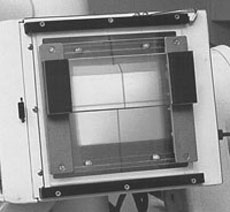
The thick part of such a wedge filter is designed to limit the amount of x-ray reaching a thinner (or less dense) body part, while the full intensity of the x-ray beam is allowed to reach the thicker body part. The filter is placed in front of the collimator (via a special adjustable filter holder, magnets, or even velcro) after collimating appropriately, because in most cases the filter is opaque and obliterates the light beam from the collimator. The gradual wedge design of the filter blends the difference between thick and thinner attenuation so that no horizontal filter line shows on the finished radiograph.
Attempts to compensate for varying thicknesses in body parts should never be made by utilizing split or gradient intensifying screens or by blocking a portion of a screen, as these improper procedures would produce the compensation after the full amount of radiation had already passed through the patient's body. Wedge compensation occurs before the radiation reaches the patient, thus providing the dual benefit of protecting the patient while obtaining a higher quality film.
Compensating filtration should be used in the following circumstances:- For all AP full spine projections
Measure and compute technique for the thickest part of the abdomen and place the thick part of the filter in front of the thinner thorax. This avoids full spine films that are too dark at the top and too light through the pelvis at the bottom. (Lateral full spine projections should be avoided; rather, three separate sectional views should be produced to accompany the APFS projection [plus one additional AP cervical film; either APOM or 15° tilt-up].)
- For virtually all AP thoracic projections
Procedure and rationale same as above. (The only exception to using a filter on an AP thoracic projection would be for a male patient with unusually developed pectoral muscles (as a body builder) and a very thin waist.)
- For most lateral thoracic projections
Measure side-to-side across the shoulders, on the outside of the arms, and compute technique sufficient to penetrate that thick and dense region. Then place the wedge filter inverted so that the thick part of the wedge attenuates the x-ray beam through the lower air-filled thorax, and the full amount of the beam is available to penetrate the dense upper thorax. This usually facilitates visualization of the difficult-to-see upper thoracic vertebrae without overpenetrating the lower thoracics.
- For most lateral lumbar projections on females
Measure side-to-side at widest part of hips and compute technique sufficient to penetrate that. Then place the wedge filter so that the thick part of the wedge attenuates the x-ray beam through the thinner waist, and the full amount of the beam is available to penetrate the dense pelvis. This avoids lateral lumbar films that are too dark at the top while being underpenetrated at the lumbosacral junction.
-
For all AP and oblique foot projections
Measure the thickest part of the foot, up near the ankle. Place wedge in front of the forefoot, thus avoiding overpenetration of the toes, while allowing visualization of the tarsals.
- For all swimmer's projections
Place filter to attenuate the beam through the cervical spine above the clavicle, while allowing visualization of the cervicothoracic junction.
- For neutral AP shoulder projections
Avoids overpenetrating the acromioclavicular joint while allowing visualization of the glenohumeral joint.
See her website at: http://www.drvxray.com/